Pacemaker
A pacemaker, also known as an artificial cardiac pacemaker, is an implanted medical device that generates electrical pulses to regulate the heartbeat. It is used to treat conditions where the heart's natural electrical system provides an inadequate or irregular rhythm, such as bradycardia or heart block. Modern pacemakers are programmable, allowing cardiologists to tailor therapy to individual patient needs, and advanced models can coordinate the contractions of multiple heart chambers to treat heart failure. For Canadians, pacemakers represent a critical and widely used medical technology that sustains cardiac function and improves quality of life for hundreds of thousands of patients.
Methods of Cardiac Pacing
Pacemakers can be applied through several methods, ranging from temporary emergency procedures to permanent surgical implantation. Percussive pacing is an old, life-saving technique involving striking the lower sternum with a closed fist to mechanically induce a ventricular beat until an electrical device is available. The British Journal of Anaesthesia notes the strike must raise ventricular pressure to 10-15 mmHg to induce electrical activity. Transcutaneous pacing (TCP) is a common emergency procedure where external pads on the chest deliver electrical currents to stabilize significant bradycardias of all types, serving as a bridge to more definitive therapy like transvenous pacing. For patients undergoing open heart surgery, temporary epicardial pacing leads may be placed directly on the heart's outer surface (epicardium) to maintain satisfactory cardiac output should the procedure create an atrioventricular block.
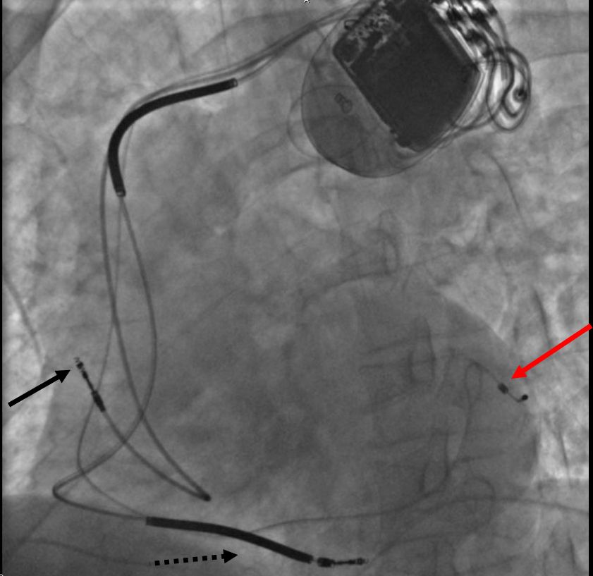
Temporary transvenous pacing involves threading a wire through a vein under sterile conditions into the right atrium or right ventricle, connecting it to an external pacemaker outside the body. This is often used as a bridge until a permanent pacemaker is implanted or until pacing is no longer needed. The standard for long-term treatment is permanent transvenous pacing, where one or more electrode leads are passed through a vein, through the heart's valves, and positioned within a chamber, facilitated by fluoroscopy for visualization, before being connected to a generator implanted under the skin near the collarbone. A significant advancement is leadless pacing, where a capsule-sized generator is implanted directly inside the heart using a steerable catheter fed into the femoral vein via a groin incision, eliminating the need for separate pacing leads and their associated long-term failure risks. Permanent epicardial pacing leads are an alternative surgical option, where leads are sewn or screwed onto the heart's surface and tunneled to the generator pocket.
Permanent Pacemaker Types and Basic Function
Permanent pacemakers are classified by the number of heart chambers they pace and their operating logic. A single-chamber pacemaker uses one lead placed in either the atrium or the ventricle. A dual-chamber pacemaker uses leads in both the atrium and ventricle, helping to coordinate the timing between these chambers to mimic natural heart function more closely by assisting the heart in coordinating function between the atria and ventricles. A biventricular pacemaker, used for cardiac resynchronization therapy (CRT), employs three leads: one in the atrium and one in each ventricle to improve the synchronization of ventricular contractions and is more complicated to implant. Rate-responsive pacemakers incorporate sensors that detect changes in the patient's physical activity, often via an accelerometer, and automatically adjust the pacing rate to fulfill the body's metabolic needs.
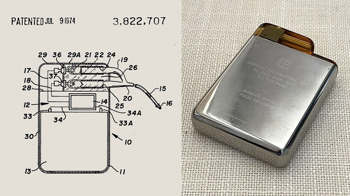
The core function is demand pacing, where the device monitors the heart's native electrical activity and only delivers a pulse if it does not detect a natural beat within a set period, commonly one second. The generator is a hermetically sealed device containing a lithium-iodide battery, a sensing amplifier to process natural heart beats, computer logic, and output circuitry to deliver the pacing impulse. Common programmable modes include VVI or VVIR for ventricular demand pacing, suitable when no atrial synchronization is needed as in atrial fibrillation, and DDDR for dual-chamber rate-responsive pacing, which covers a wide range of conditions though it requires separate atrial and ventricular leads and careful programming. The device must reliably 'capture' the heart muscle, requiring a minimum electrical voltage and pulse width known as the threshold. The actual voltage used is typically 50 to 100 percent higher than this minimum to ensure reliability. Sensing, or detecting intrinsic heart activity, is governed by sensitivity settings. Too low sensitivity causes over-sensing of P waves, T waves, or noise, while too high sensitivity results in under-sensing and missed beats.
- Single-Chamber: One lead in atrium OR ventricle.
- Dual-Chamber: Leads in both atrium AND ventricle for coordinated timing.
- Biventricular: Three leads for CRT (atrium, right ventricle, left ventricle).
- Rate-Responsive: Adjusts rate based on sensors (e.g., activity).
- Common Modes: VVI/VVIR, AAI/AAIR, VDD, DDDR.
- Generator Components: Battery, sensing amplifier, computer logic, output circuitry.
- Key Parameters: Pacing threshold (voltage/pulse width), sensing sensitivity.
Cardiac Resynchronization Therapy and Conduction System Pacing
Cardiac resynchronization therapy (CRT) is a specialized application of biventricular pacing for patients with heart failure and ventricular dyssynchrony, where the left and right ventricles do not contract simultaneously, a condition affecting approximately 25 to 50 percent of heart failure patients. A biventricular pacemaker paces both the septal and lateral walls of the left ventricle to coordinate contractions. Standard CRT devices (CRT-P) use at least two leads: one passes through the vena cava and right atrium into the right ventricle to stimulate the septum, and another passes through the vena cava, right atrium, and coronary sinus to pace the epicardial wall of the left ventricle. A third lead in the right atrium is often added for patients in normal sinus rhythm to facilitate synchrony with atrial contraction. For patients also at risk of life-threatening arrhythmias, a combined device (CRT-D) includes implantable cardioverter-defibrillator functions to provide protection against life-threatening arrhythmias. Clinical trials have shown CRT reduces mortality and improves quality of life in eligible patients with heart failure symptoms, a left ventricular ejection fraction less than or equal to 35 percent, and a QRS duration on electrocardiogram of 120 milliseconds or greater.
Conventional pacing from the right ventricular apex can have detrimental long-term effects, including increased risk of atrial fibrillation, heart failure, weakening of the heart muscle, and potentially shorter life expectancy. Conduction system pacing techniques like His bundle pacing (HBP) and left bundle branch area pacing (LBBAP) aim to stimulate the heart's natural electrical conduction pathways directly, leading to a more natural or perfectly natural ventricular activation. By stimulating the His-Purkinje fiber network directly with a special lead and placement technique, HBP causes a synchronized and therefore more effective ventricular activation and avoids long-term heart muscle disease. In some cases it can also correct bundle branch block patterns. These methods represent a significant shift in pacing strategy to avoid the negative consequences of traditional site pacing and have generated strong research and clinical interest.
Surgical Implantation and Maintenance
Pacemaker implantation is typically a procedure lasting 30 to 90 minutes, performed under local anesthetic with sedation or under general anesthetic. An antibiotic is administered to reduce infection risk, and the skin is prepared by clipping hair and cleaning with a disinfectant like chlorhexidine. The surgeon makes an incision below the collarbone and creates a pocket under the skin, usually just above the pectoralis major muscle (prepectoral), though sometimes beneath the muscle (submuscular), to house the titanium generator. Using fluoroscopic X-ray guidance, the leads are threaded through a large vein, such as the subclavian or cephalic vein, and positioned within the appropriate heart chambers: the right ventricle, right atrium, or coronary sinus, depending on the pacemaker type. Following surgery, patients must keep the wound clean and dry until healed, and avoid certain shoulder movements within the first few weeks to prevent lead dislodgement.
Pacemaker batteries typically last 5 to 10 years. When battery depletion nears, the generator is replaced in a procedure that is usually simpler than the initial implant. The existing device is removed through an incision, the leads are disconnected from the old generator and reconnected to a new one, and the new device is inserted into the existing or a new pocket. After implantation, the device requires periodic checkups to ensure it is operating correctly and the battery is functional. These checks can be performed remotely via telemonitoring or during in-person clinic visits where a cardiologist can select optimal pacing modes and adjust settings. The outer casing of pacemakers is designed to be rarely rejected by the body's immune system and is usually made of titanium, which is inert in the body.
- Anesthesia: Local with/without sedation, or general.
- Pocket Location: Prepectoral (above chest muscle) or submuscular.
- Lead Targets: Right ventricle, right atrium, coronary sinus.
- Imaging: Fluoroscopy guides lead placement.
- Post-op Restriction: Avoid strenuous arm/shoulder movement for weeks.
- Generator Material: Titanium casing.
- Replacement: Generator swapped every 5-10 years. Leads often reused.
- Follow-up: Periodic checks for function, settings, and battery status.
Technological Advancements and Programming
Pacemaker technology has evolved significantly from fixed-rate devices to sophisticated, microprocessor-controlled systems. A major advancement is rate-responsive pacing, where sensors detect parameters like physical activity via an accelerometer, body temperature, dissolved oxygen or carbon dioxide levels (pO2-pCO2), the QT interval, ATP levels, or adrenaline to dynamically adjust the pacing rate to metabolic demand. The first dynamic pacemaker was invented by Anthony Rickards of the National Heart Hospital in London, UK, in 1982. Modern dual-chamber devices not only pace but also carefully time atrial and ventricular contractions to improve pumping efficiency, which is particularly beneficial for some patients with congestive heart failure, and they can keep the amount of right ventricle pacing to a minimum to prevent worsening of heart disease.
Programming advancements now allow clinicians to minimize unnecessary right ventricular pacing, which studies like the DAVID trials have linked to exacerbating heart failure and increasing the incidence of atrial fibrillation. The transition to microprocessor control has enabled complex algorithms for managing atrioventricular delays, mode switching, and dynamic compensation for both actual and anticipated respiratory loading. Future directions may involve further integration with physiological sensors and the application of such dynamic control systems in artificial heart technology. Advances in transitional tissue welding and stem cells may also impact future device integration and artificial organ development. The ability to externally program the devices allows cardiologists to non-invasively fine-tune pacing modes, rates, and sensitivity long after implantation to adapt to changing patient conditions.
| Advancement | Key Feature | Purpose/Benefit |
|---|---|---|
| Rate-Responsive Pacing | Sensors (e.g., accelerometer) | Adjust heart rate to metabolic demand during activity |
| Microprocessor Control | Programmable logic | Enables complex pacing algorithms and mode switching |
| Dual-Chamber Timing | Coordinates atrial & ventricular contraction | Improves pumping efficiency, mimics natural rhythm |
| Minimal RV Pacing Algorithms | Reduces unnecessary ventricular pacing | Prevents worsening heart failure & atrial fibrillation |
| Leadless Technology | Capsule generator in heart | Eliminates lead failure risks, simpler implantation |
| Conduction System Pacing | His bundle or left bundle area pacing | More physiologic activation, preserves long-term function |
Clinical Indications and Patient Considerations
Pacemakers are primarily indicated for conditions where the heart's electrical system fails to maintain an adequate rate or rhythm. This includes sick sinus syndrome or sinus node disease, where the heart's natural pacemaker is unreliable, and various degrees of atrioventricular block (AVB), where electrical signals between the atria and ventricles are impaired. The choice of pacemaker type depends on the specific conduction abnormality. For example, AAI or AAIR mode is suitable when atrioventricular conduction is intact but the sinoatrial node is faulty, while VDD mode can manage AVB by sensing the atrial beat and triggering a ventricular beat after a normal delay of 0.1 to 0.2 seconds. For patients with heart failure and ventricular dyssynchrony, a biventricular pacemaker (CRT-P or CRT-D) is indicated to resynchronize contractions, with eligibility often based on ejection fraction and QRS duration.
Device selection and programming require careful consideration of individual patient physiology and the underlying cardiac pathology. Factors like lead impedance, which affects current drain and battery life, and the integrity of the electrode-tissue interface are critical for long-term performance. The pacing system must be configured to avoid complications such as phrenic nerve stimulation or inappropriate mode switching. Patients with pacemakers must be aware of certain environmental interactions, though modern devices have filters to protect against most everyday electromagnetic interference. They should inform medical and dental professionals about the device before procedures. The overall goal is to use the most appropriate device and settings to alleviate symptoms like fatigue and dizziness, prevent complications like syncope, and improve longevity and quality of life, with ongoing follow-up to ensure optimal function.
Pacing Modes and Electrical Characteristics
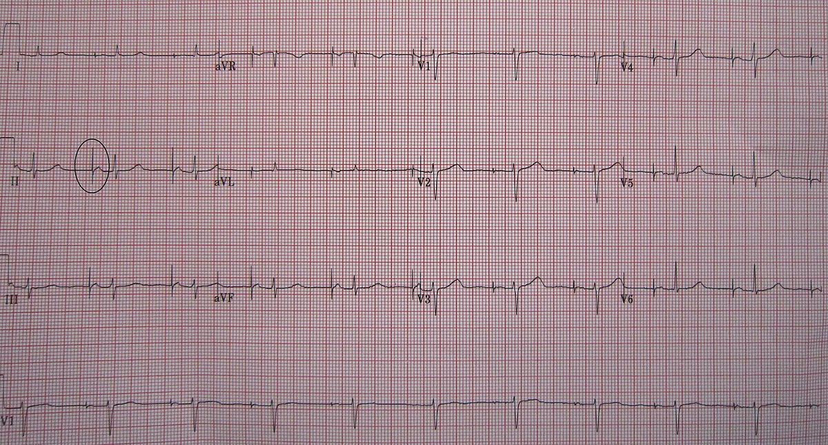
The operating mode of a pacemaker is described by a standardized code that indicates which chambers are paced, which are sensed, the response to sensing, and rate modulation. In a ventricular-demand pacemaker (VVI), the device paces and senses in the ventricle and inhibits pacing when it senses intrinsic activity, producing a narrow vertical spike on the ECG just before a wide QRS complex. An atrial-demand pacemaker (AAI) paces and senses in the atrium, with its spike appearing just before the P wave. Triggered modes, such as a ventricular triggered pacemaker, deliver an impulse immediately after sensing intrinsic activity in that chamber, appearing as a spike simultaneous with the QRS complex. The more comprehensive DDDR mode paces and senses in both the atrium and ventricle, can trigger or inhibit responses, and includes rate modulation, making it versatile but requiring complex programming for optimal results.
The electrical characteristics of the pacing system are fundamental to its operation. The pacing threshold is the minimum voltage and pulse width needed to reliably capture the myocardium and cause contraction. The programmed output is set higher than this threshold as a safety margin. The pacing circuit can be unipolar, where the cathode is at the lead tip and the anode is the pacemaker generator case, or bipolar, where both cathode and anode are within the lead. Sensing sensitivity determines the minimum intracardiac voltage the device will recognize as a native beat. Setting it correctly is crucial to avoid over-sensing (e.g., of T-waves) or under-sensing (missing P-waves or QRS complexes). These parameters are tested at implantation and during follow-up checks to ensure the system delivers therapy only when needed and does not misinterpret extraneous signals.
Questions about Pacemaker
What is a pacemaker and what does it do?
A pacemaker is an implanted medical device that generates electrical pulses to regulate the heartbeat. It is used when the heart's natural electrical system produces a rhythm that is too slow, too fast, or irregular. The device monitors heart activity and delivers pulses to cause the heart chambers to contract and pump blood effectively, thereby maintaining an adequate heart rate and improving cardiac output. Modern devices are programmable and can adapt pacing based on the body's needs.
How is a pacemaker implanted?
A pacemaker is typically implanted in a procedure lasting 30 to 90 minutes. Under local or general anesthetic, an incision is made below the collarbone. The generator is placed in a pocket under the skin or muscle, and insulated wires (leads) are threaded through a vein into the heart, guided by X-ray imaging (fluoroscopy). The leads are connected to the generator, which is then secured in the pocket before the incision is closed. Antibiotics are given to reduce infection risk.
How long does a pacemaker battery last?
The lithium-iodide battery in a pacemaker generator typically lasts between 5 and 10 years. Battery life depends on factors like how often the device paces the heart and its programmed settings. When the battery nears depletion, the generator is replaced in a surgical procedure where the old unit is removed and a new one is connected to the existing leads. This replacement is usually simpler than the original implantation.
What is a biventricular pacemaker?
A biventricular pacemaker is a type of pacemaker used for cardiac resynchronization therapy. It has multiple electrodes stimulating different positions within the ventricles to improve their synchronization. This device is used for people with heart failure in whom the left and right ventricles do not contract simultaneously. To achieve this, it paces both the septal and lateral walls of the left ventricle. Biventricular pacing alone is referred to as CRT-P for pacing.
What are the risks of pacemaker surgery?
As with any surgery, pacemaker implantation carries risks including infection, bleeding, bruising, and allergic reactions to anesthesia. Specific to the procedure are risks of pneumothorax (collapsed lung), lead dislodgement, or perforation of the heart muscle or blood vessels by the leads. There is also a risk of device malfunction or interaction with strong electromagnetic fields. Patients are advised to avoid certain arm movements shortly after surgery to minimize the risk of lead displacement.
Can you live a normal life with a pacemaker?
Yes, most people with pacemakers can resume normal activities, including exercise, work, and travel. Certain precautions are advised, such as informing medical and dental professionals about the device before any procedures and being aware of potential interference from strong electromagnetic sources like industrial equipment or certain security systems. Regular checkups are needed to monitor device function and battery life. The device itself is designed to be durable and compatible with the body.
Sources and further reading
- Pacemaker (Wikipedia)
- A sub-study of the LANDMARK trial (pubmed.ncbi.nlm.nih.gov)
- Pacemaker (ottawaheart.ca)
- Implantable pacemaker | Heart and Stroke Foundation (heartandstroke.ca)
Checked by the Your Space Hamilton desk. Last updated 2026-10-20.